September 2, 2013 - by Simone Ulmer
It is common knowledge that mechanical stress on bones, arising through activities such as jogging or walking, encourages bone formation. However, it has remained unclear how exactly the bone-forming cells in the bone marrow respond to mechanical stimuli – or how their bone-resorbing counterparts respond in the absence of strain. Now, researchers from ETH Zurich have demonstrated for the first time how local mechanical stress and bone formation or resorption (bone breakdown) are linked by combining lab experiments and simulations on CSCS supercomputer “Monte Rosa”. These have revealed that eighty per cent of the build-up and breakdown of bone substance is controlled by mechanical stimuli.
Strain encourages balanced bone formation
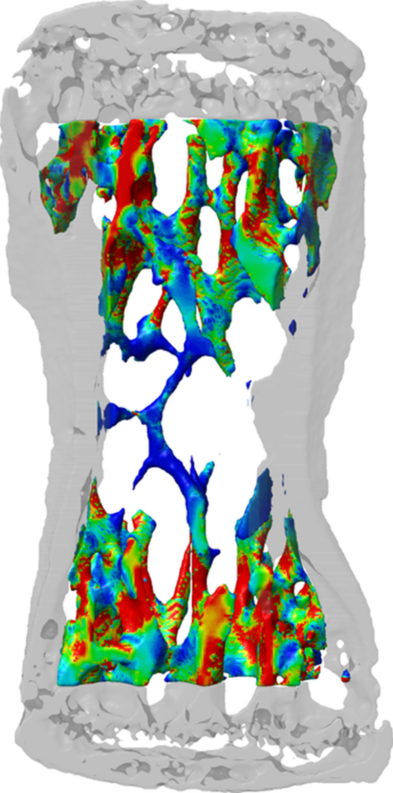
The simulations reveal how the mechanical strain exerted locally in the lab – by stretching a vertebral body in a mouse tail – leads to the build-up of the bone substance in certain places and its resorption elsewhere. According to the researchers, the results also confirm the assumption that bone substance is formed where it is needed and resorbed where it is not. A well-balanced interplay between osteoblasts, which are responsible for bone formation, and osteoclasts, which resorb and break down bones, is important for a healthy organism. Both the over- and underproduction of these two kinds of cells lead to abnormal changes in the bone structure. “Our study is therefore essential for gaining a better understanding of bone diseases and how to develop new medications,” says Ralph Müller, who is the study director and a professor of biomechanics at ETH Zurich.
The scientists used different groups of mice for their experiments. While a certain caudal vertebra segment was stressed mechanically three times a week for a period of four weeks in one group, this part of the experiment was omitted for the control group. Moreover, there was another test group in which the animals’ ovaries were removed. The resulting lack of oestrogen has a negative impact on bone formation and leads to bone resorption – presumably one of the reasons why around thirty per cent of women develop osteoporosis after the menopause.
Experiment and simulation indicate correlation
During the experiments, the researchers regularly took high-resolution computed tomography images of the animals. Superimposed and processed into three-dimensional representations using imaging processing, the images produced spatial models displaying the areas of bone formation, bone resorption and areas where nothing had changed. The scientists then exposed these models virtually to different forces on the supercomputer and calculated what would happen next for every area. Through these simulations, the researchers were able to study the link between local mechanical stress and its impact on the bone at the cellular level. “The results clearly show that the activity of both the osteoblasts and the osteoclasts is controlled by mechanical strain,” says Ralph Müller. “High local strains lead to the formation of the bone and low strains provoke its resorption.” According to the researchers’ analyses, the probability of bone substance being resorbed under increasing mechanical strain decreases exponentially while bone formation increases exponentially. Moreover, bone resorption appears to be controlled mechanically to a considerably higher degree than bone formation, especially in mice without ovaries, where non-specific bone resorption increases significantly in the absence of mechanical strain. In other words, bone substance is also resorbed in places where it is actually needed.
For the scientists, the results echo the mounting evidence that oestrogen receptors are involved in the bone-cell response to mechanical stimuli. In short: a lack of oestrogen receptors limits targeted bone resorption.
Reference
Schulte FA et al.: Local Mechanical Stimuli Regulate Formation and Resorption in Mice at the Tissue Level, PLoS ONE (2013), 8(4): e62172. Doi:10.1371/journal.pone.0062172
